Friday, July 30, 2010

Glue Traps

Sadly, many of the animals we treat in the clinic are there because human-induced injuries. One of the most frustrating are glue traps. Glue traps are pieces of cardboard with a sticky gel on top that is marketed as a rodent trap. What happens is the rodent runs across it and becomes stuck to the gel. The rodents are then stuck there to die of starvation and many have been known to chew their legs off in desperation to get away.

The whole concept of the glue trap in regards to rodents is inhumane; it is even more frustrating when other species become stuck to these traps. Songbirds often come in stuck to these traps. If they are alive when they get to us, they require several baths in alternating baths of vegetable oil (to release them from the glue and wash off feather contamination) and dawn dishsoap (to wash the oil off) . This process is very stressful for the bird.

The other day 3 bats, a big brown and two nothern (keen's) myotis, were brought in having been stuck to a glue trap. As we examined the bats, we were very saddened to see the destruction glue traps can do. All three bats had fractured bones and wing-webbing ripped off. The bones were amputated (not attached to the bats) and once wing webbing is torn, it cannot be repaired. These injuries necessitated humane euthanasia.

We can only hope that those who read this post will pass on the knowledge of dangers of glue traps, in hopes that people will stop using them.

I'm going to refrain from posting pictures as some may find them graphic.

Friday, July 23, 2010

babies!


Baby season is still in full force so I thought I'd share some pictures.


These are cedar waxwings who will be fledging soon. All songbirds go through a stage call the fledgling stage where they jump out of the next and spend up to a week on the ground learning how to feed themselves and fly. Their parents are still around, feeding them on the ground, but less often (tough love;) and won't come around if you are outside watching the babies. This is why we get so many fledglings into the center--people think they are abandoned when mom and dad are simply waiting for the predators (aka humans) to go away so they can feed their babies again.

We know these are cedar waxwings b/c the mouth color is a hot pink with iridescent strips on the sides (much more striking in person!)


Compare that to these nestling house sparrows. Not all baby birds look this different though. The younger they are, the harder they are to tell apart and sometimes we simply have to wait until they grow up until we can ID their species.


Since fledglings are like "teenagers", they aren't the smartest lot.
This fledgling/juvenile piliated woodpecker came in with hindlimb weakness/tail paralysis (likely from spinal trauma) and a foreign body in his stomach (see xray below--the white sharp edged trapezoid towards the right/bottom is a piece of glass/plastic). I had to go in surgically and remove it from his stomach. He recovered from his surgery well, and is VERY slowly regaining movement in his legs. I'm hoping he'll fully recover so we can release him, but his tail needs to follow suit...


This is a young common nighthawk (VERY cool species--sort of like a large chimney swift- they eat only when flying through the air catching bugs and their legs are positioned to cling to walls-->so they can't stand when on the ground...weird). He was doing well in the nursery until he became very bloated one evening. Xray revealed he had 10-11 stones in his stomach that were obstructing him. He needed emergency surgery. Unfortunately, he died during surgery:( he was very sick, so it wasn't surprising he didn't make it.


Below are some baby opossums inside their mother's pouch! They are about the size of a dime when they are born; they climb into her pouch and latch on to a teat. Opossums have 13 nipples so can accomodate many young.


The picture below is a grey squirrel a few days old. He weighed in at ~13 grams!

Whew! that's enough for now! I'll post more pics next week:)

Saturday, July 3, 2010

updates and a new toad

the snapper has recovered from her surgery and is doing ok right now. I'm hoping we can ward off infection with antibiotics and in a few months she'll be ready for release.

the toad unfortunately didn't have as good of an outcome.

she recovered well from that surgery (the stomach healed very nicely and she felt well enough to eat a grub on her own!) but she remained lethargic and didn’t pass any stool. To determine why she wasn’t fully recovered yet, we performed a barium study. A barium study is where you feed an animal a liquid that shows up bright white on xrays. Serial xrays are taken and you can see the barium as it passes through various parts of the gastrointestinal tract. Here is one taken ~ 1 hour after we fed her the barium. The blob on the right is barium in her stomach and you can see it snaking through the intestines to the left.sn852795.JPGwe continued to take xrays but after 24 hours the barium became stuck at the junction of the small intestine and colon.sn852805.JPGShe needed surgery again or she would die. So we went to surgery but unfortunately, the part of the intestine that had all the barium in it was extremely fragile and burst. She would have needed the fragile part of her intestine cut out and the two ends sewn together–but we couldn’t find why she became obstructed in the first place. It was decided to humanely euthanize her as the surgery she needed was very risky (high rate of failure) and we couldn’t guarantee she wouldn’t become obstructed again. it seemed unfair to put her through more pain when we couldn’t give her a good prognosis.

However, admist the poor outcomes are always several more happy and healthy ones!

Monday, June 28, 2010

common snapping turtle


This snapping turtle came in with several shell fractures. Several on the top shell (carapace) which I repaired by drilling holes in the edge of the shell on either side of the fracture and looping wire through the holes to hold the fracture in place....and one big on on her bottom shell (plastron). Here is a pic of her on her back (under anesthesia)


and a close-up of the fracture....the arrow is pointing to it and the black line is parallel to it so you can see how long it is. It extends from one edge of the plastron to the other so essentially, the 2 edges of the fracture are moveable and keep moving up and done (dorsal and ventral) as it's broken all the way through in that area.
I wanted to try a new technique where I drill screws into the shell and connect the screws with wire but there were a few problems
-the fracture pieces kept collapsing dorsally (into the turtle) even with the wire/screws so if i just did that, the pieces wouldn't stay aligned....they would "V" down into the turtle
-once the screws and wire were in place, i didn't want pressure on the screws or the fracture, and once the turtle was upright (sternal) she would be putting all of her weight on the screws since they would be sticking out a bit (they don't make stainless steel screws smaller than 1/2", ugh).
-the fracture edges were VERY hard to align even when manually holding them together--they didn't want to go into their "natural" position which would make repair difficult

SO after running around the center trying to think of an answer, i came up with this...i found a plastic lid from a heavy plastic jar and drilled holes in it, then placed the screws through the holes and into the holes I had drilled into the shell--this prevented the fracture edges from collapsing into her body...however, since the lid had to be very rigid, it was difficult to hold the fracture in place while placing the lid, so the fracture didn't align correctly BUT it's not collapsing into her body; it will heal---will just take longer.

the next problem was that i didn't want pressure on the lid at all or it might bump around the fracture.

so i took one of those pool noodles, cut it in half and glued some thick pieces around the lid...
and then glued a frisbee onto the pool noodles


she woke up very slow from anesthesia and I'm afraid may not make it. Manipulating the fracture so much (b/c it was so difficult to align) and having her on her back for ~30 min may have been too stressful for her....we'll see over the next few days.


Wednesday, June 23, 2010

I love my job:)


I love my job:) I'm working 12-14 hour days, examining, admitting and determining treatments for 70+ new patients each day, rechecking the bulk of my 50+ existing cases each day, answering the phone and talking to the public when we don't have a front desk volunteer, examining nursery animals as they become ill and dealing with whatever curve balls come my way. I'm constantly multitasking and doing things i've never done before.

For instance, here is one story for tonight.
This American Toad presented very sick–she was very lethargic and her whole body was swollen. When amphibians (frogs and toads in particular) get a swollen body, it’s termed “edema disease” which can be caused by a long list of diseases–kidney disease, liver disease, electrolyte imbalances, and pretty much anything that can make an amphibian sick can cause edema disease.

A radiograph (pictured) showed a large amount of a mineral substance (likely sand/grit/stones) in her stomach (the white blob in the middle of the xray).


This grit was obstructing her stomach, not allowing anything to pass. The only way to get it out is surgery. This toad was so sick that she would likely die during surgery, but she would definitely die if we didn’t get the obstruction out of her stomach soon.So she went to surgery. By cutting through her skin and muscle layers, I was able to see the stomach, cut it open, pull the grit out and sew her back up. She survived surgery but is still very sick and may die despite our best efforts.We aren’t sure why she got the obstruction. Toads live in an environment where sand and grit are abundant–why did she eat so much to get obstructed? She was kept as a pet (wild animals never make good pets) so there is the possibility that having a limited environment and diet, she accidentally ate sand looking for food, or her insects were covered in a large amount of dirt–or perhaps she has some gastrointestinal disease that caused her stomach motility to slow, allowing small amounts of sand to accumulate over time–we’ll never know what exactly caused it. We’re just hoping we got it out in time and she’ll pull through.


Friday, May 21, 2010

some cases of old...


thought these might be fun to post until i start my new job and have new cases to talk about:)

A small mature dog came in having been hit by a car. He was quite shocky so we immediately started IV fluids and pain medications. Owners approved xrays right away and here's what we found.
It's a bit hard to see in this small picture, but at the tip of the arrow are several fractures of the hip bones.
When animals are hit by cars, they can go into shock for several reasons. This guy was incredibly painful b/c of the fractures--this and the trauma in general can cause shock. He needed to stay in the hospital on IV fluids to help get him out of shock and continued pain medications to make him more comfortable. Also, many problems don't arise until several hours or even a day after the trauma--things like lung bruising may not be evident on the xrays until the next day so it's VERY important to keep these guys in hospital for treatment AND continued monitoring. Eventually (once he's more stable-->either at the regular vet or a referral center) he might need surgery to repair some of these fractures.

After discussing all of this with the owners, they explained they had no money. They knew how much xrays would cost when they approved it, but couldn't really afford that. I gave the owners the option of euthanizing which they refused. So they took him home were going to try to find a low-cost clinic in the morning. I tried to give him as much pain medication before he left as possible, but there was a good chance he would die at home. very sad. and my hands were tied.

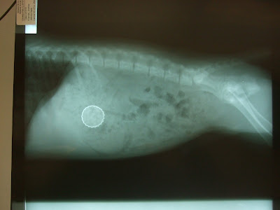
A 20 lb puppy came in for 'possibly' eating a bottle cap. The owner saw the bottle cap on the floor and a few minutes later, it was gone--no where to be found. The puppy was acting completely normal when it came in. We took some xrays...below...

Definitely a bottle cap in the stomach- and compared to the size of the puppy, it's pretty big! Would likely obstruct the small intestine if we let it stay there.

So I gave the owner two options: 1) induce vomiting--risks include the sharp edges of the bottle cap scraping the esophagus on the way up-->causing scar tissue when it heals--> causing a stricture--> causing swallowing problems-->causing aspiration pneumonia +/- death...a little extreme and not a huge risk, but it's happened before. OR 2) surgery--go into the abdomen, cut into the stomach and take it out, risks include normal surgical risks (death, bleeding, infection, pain, etc) and surgery is much much more expensive (we're talking ~$300 for vomiting and $2000 for sugery...). So the owner chose to induce vomiting and voila!


The bottle cap came up along with huge amounts of dog food she just ate. Since it came up coated in partially digested food, likely no scratching of the esophagus but I sent home a medication that would help an scratches heal if they did occur.

what i should have done is sat the owner down for a serious intervention about his choice in beer-Labatt's blue? c'mon! even illinois has some decent local brews.
pft.

Wednesday, May 5, 2010

interesting article

this article talks about how veterinarians are 4x as likely as the average person and twice as likely compared to other health professionals, to commit suicide.


i just thought it was an interesting factoid--I think people usually think that being a veterinarian is less stressful than being an MD, but that may not be the case. Or perhaps the profession attracks those who are more likely? in any case, I have a lot more respect for private practitioners having been on. what a task.